
Learn about our clinic and how we help dogs and cats throughout Connecticut and the northeast US.
Meet the team
The clinic
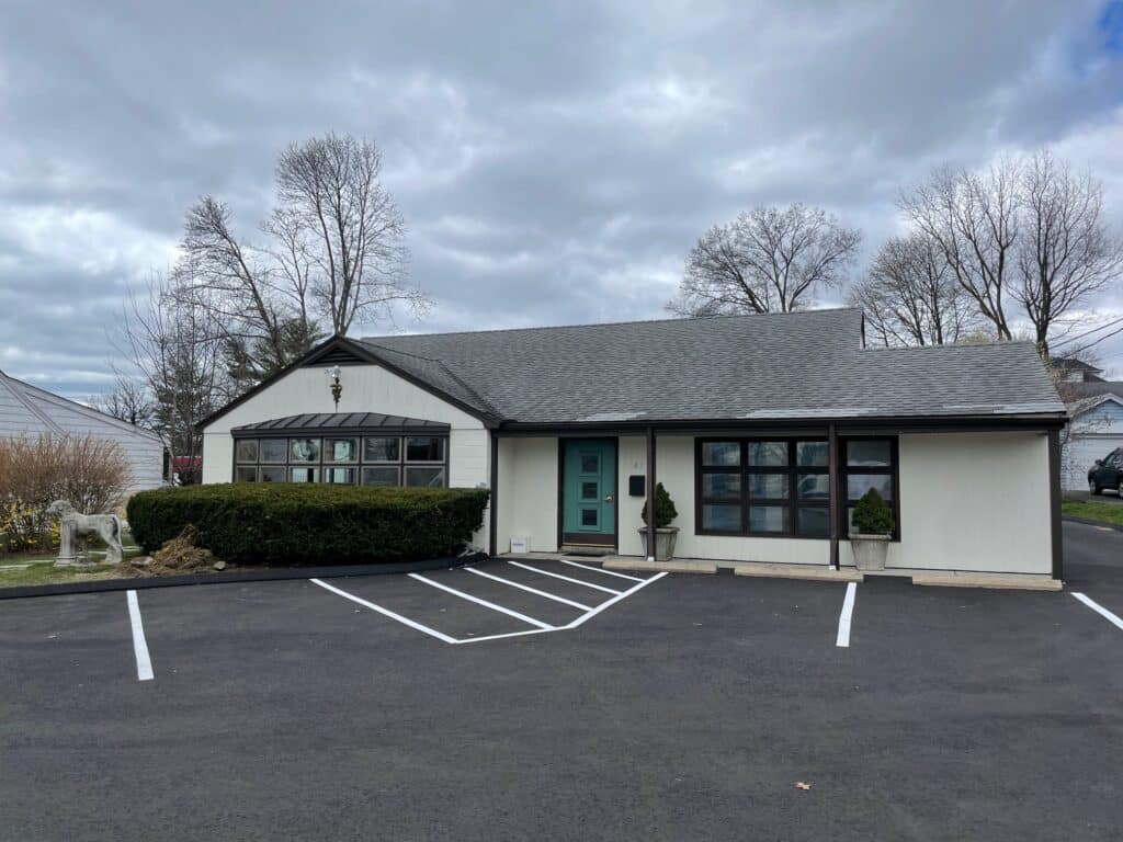
Unleashed Veterinary Dermatology is Connecticut’s only clinic that is dedicated to veterinary dermatology.
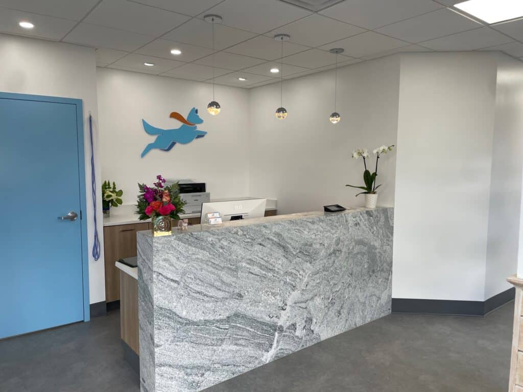
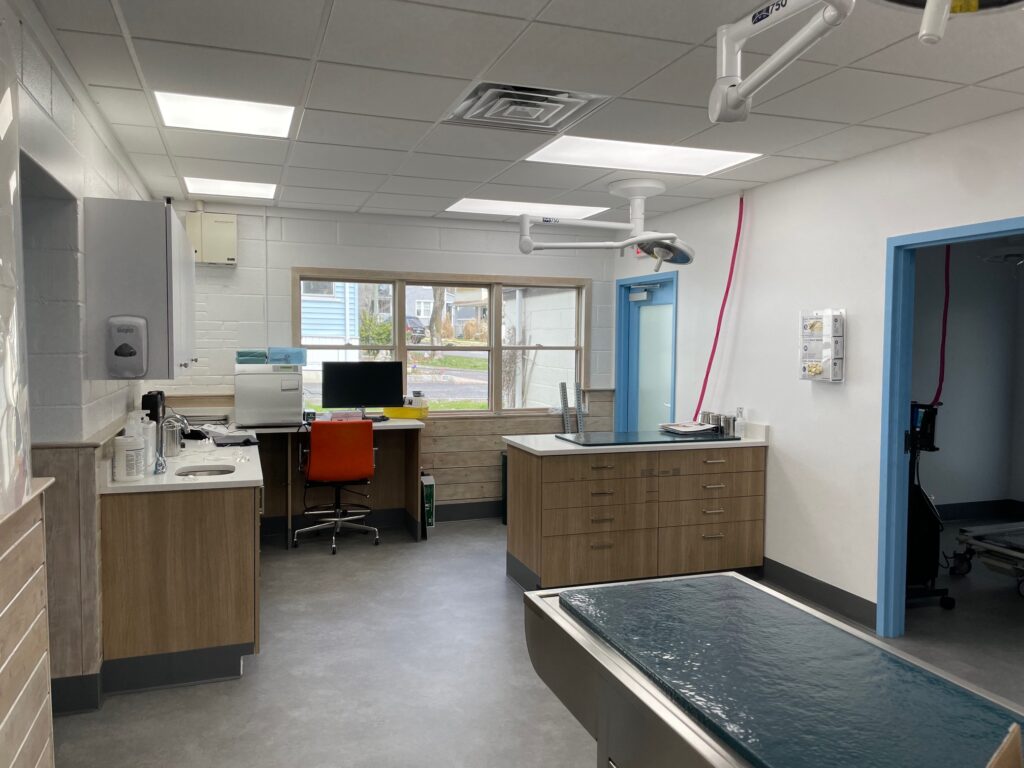
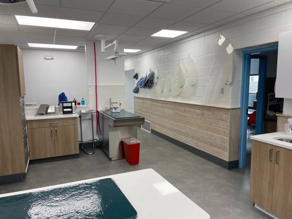
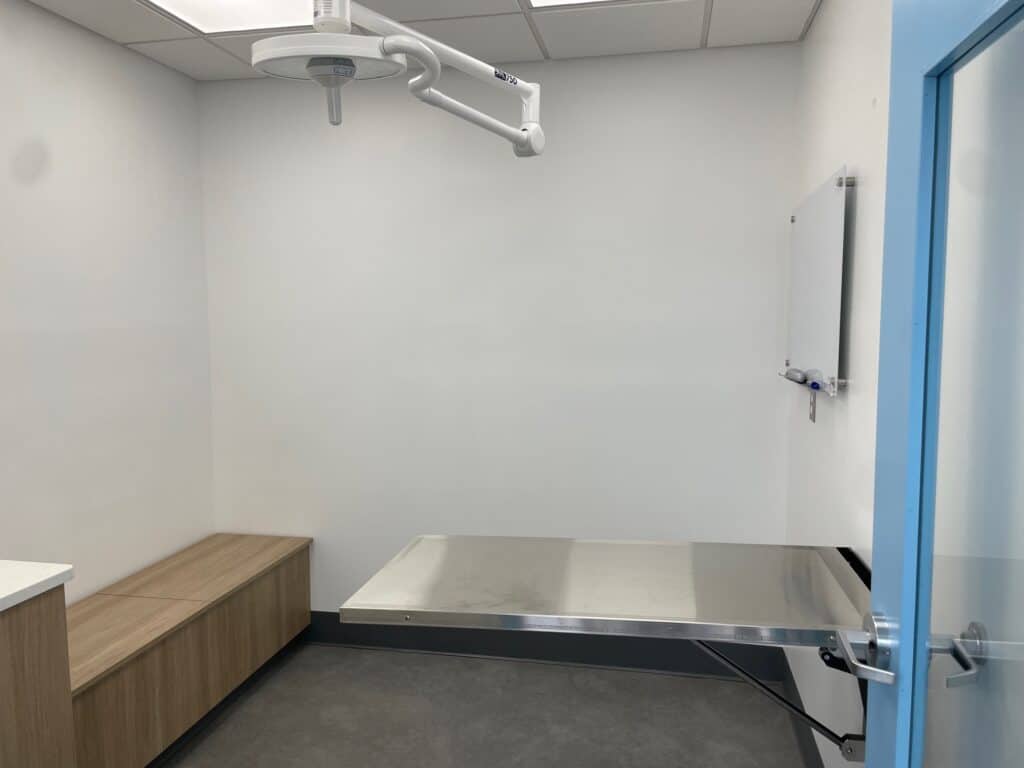
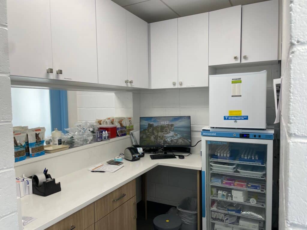
Recognizing Connecticut’s unmet need for veterinary dermatology specialists, Dr. Falk founded the practice in 2024, purchasing the former Shakespeare Veterinary Hospital from its owner, Dr. Milos. We then renovated and the clinic and equipped it to be a state-of-the-art center for veterinary dermatology. In addition to exam rooms, we have a large treatment area, a surgery suite, a laboratory, and a pharmacy.
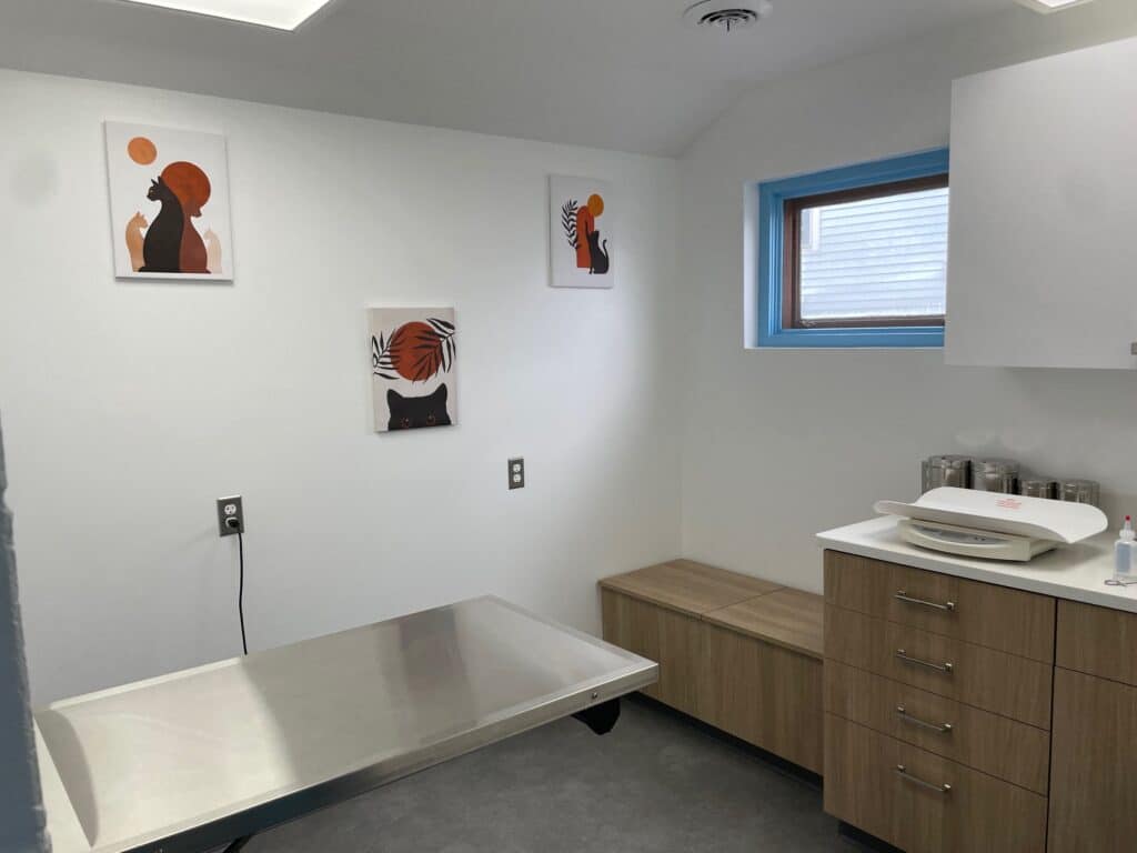
Dr. Falk is Fear Free certified, and the clinic was renovated with Fear Free principles in mind. In that vein, one of our exam rooms is dedicated solely to cats, and we will be setting up a patio outdoors that you are welcome to use in place of our waiting room if you or your pet so prefers.
In addition to Dr. Falk, our staff includes two veterinary technicians, one client care coordinator, and two veterinary assistants. We are confident that the excellence of our staff transfers through to the care that we deliver.
The clinic is just a few minutes from I-95, the Merritt, and the Stratford Metro North stop. Our primary parking lot for clients is in the front of the building. If you need a handicap accessible entrance, please park in handicap spot in the rear lot and enter through the back door, which has a ramp. If your dog has to go, there is grass in the front of the clinic and doggy pickup bags. There is a baby changing table in our new handicap-accessible bathroom.
As Dr. Falk is one of only three veterinary dermatologists in the state of CT, many of our clients come from far away, including from neighboring states like NY, MA, and RI. Whether you are coming from near or far, we welcome you to our clinic and look forward to treating your pet.
Take a tour

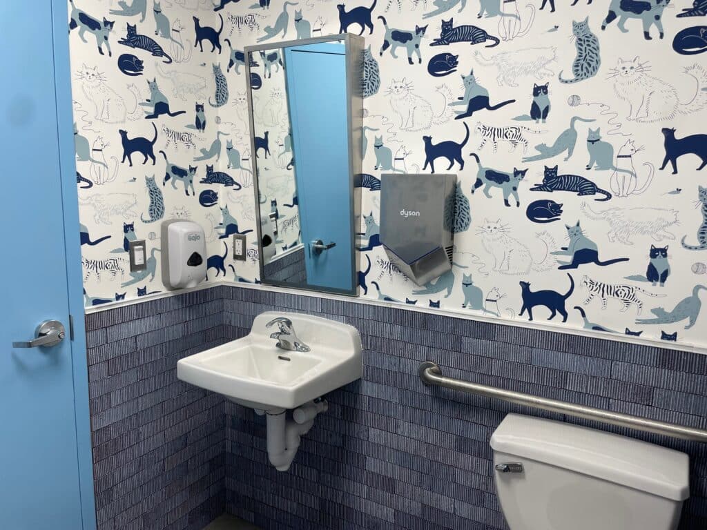
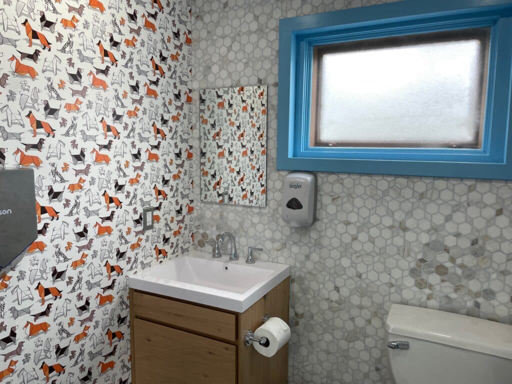
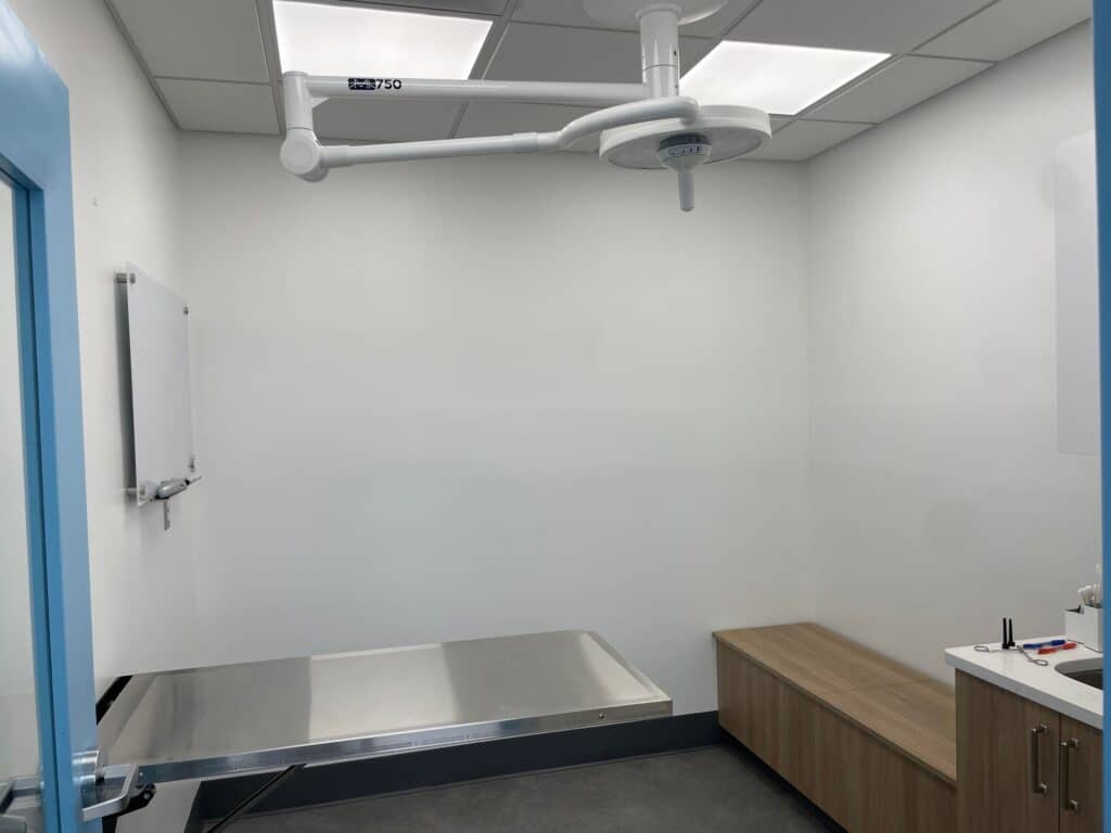

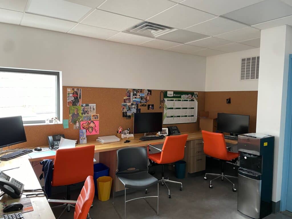
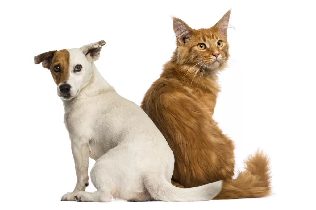
Veterinary dermatologists diagnose and treat diseases of the skin of animals. This includes a wide spectrum of diseases, ranging from allergies and ear infections to auto-immune disease and skin cancer. Skin disease can also present as a manifestation of an underlying internal disease process. Veterinary dermatologists are therefore also trained in otoscopy, immunology, allergy, and internal medicine.
Veterinary dermatologists must be board-certified by the American College of Veterinary Dermatology (ACVD). The purpose of the ACVD is to promote excellence in veterinary dermatology, oversee postgraduate training in veterinary dermatology, sponsor research, and organize scientific and educational programs for both veterinary dermatologists and general practitioners. There are currently about 300 ACVD board-certified veterinary dermatologists worldwide.
Veterinary dermatologists diagnose and treat diseases of the skin of animals. This includes a wide spectrum of diseases, ranging from allergies and ear infections to auto-immune disease and skin cancer. Skin disease can also present as a manifestation of an underlying internal disease process. Veterinary dermatologists are therefore also trained in otoscopy, immunology, allergy, and internal medicine.
Veterinary dermatologists must be board-certified by the American College of Veterinary Dermatology (ACVD). The purpose of the ACVD is to promote excellence in veterinary dermatology, oversee postgraduate training in veterinary dermatology, sponsor research, and organize scientific and educational programs for both veterinary dermatologists and general practitioners. There are currently about 300 ACVD board-certified veterinary dermatologists worldwide.
No, you may be referred or you may book an appointment anytime. The best practice is to ask your primary care veterinarian to refer your pet to us so that he/she can help us understand the history of your pet’s problems. However, whether you are referred or not, we will work closely with your primary care veterinarian.
We will be happy to assist you with your insurance claim forms. In veterinary medicine in general, you should expect to pay your full bill at the time of your visit and then get reimbursed by your insurance company. We find that most clients have good success with their claims.
If you have Trupanion pet insurance, we can also bill your insurance directly.
Our office is at 47 Nichols Ave. in Stratford, CT
After over 45 years in its location at 47 Nichols Ave., Dr. Milos retired in 2023 and sold his clinic to Dr. Falk.
Contact our office if you want a virtual visit and we will be happy to determine if a virtual visit is appropriate. In most cases, virtual visits are not preferred, because we need to examine your pet. Virtual visits cannot be used to establish a veterinary-client-patient relationship (VCPR), so Dr. Falk must have seen your pet before a virtual visit can be considered.
No, as a veterinary clinic, we do not treat people.
No, we only treat dogs and cats. A recommendation for a nearby exotics specialist is the South Wilton Veterinary Group.
The best place to buy your pet’s medications is from a veterinarian. You can order them for pickup at our clinic, buy from our online pharmacy, or buy from your other veterinarians. We offer free shipping when you buy directly from us. You are also welcome to use third-party online pet retailers, though we caution clients that these tend to be paperwork hassles that frequently involves a great deal of back and forth and delays. Some third-party pharmacies have been known to buy and sell nearly expired medications. Since we have endeavored to make our pharmacy’s prices be competitive with anything you find on the internet, and we can provide you with a streamlined ordering process, please consider buying from us (or your other veterinarians) unless you have a good reason not to.
We accept cash, checks, Visa, Mastercard, Discover, American Express, CareCredit, and Trupanion direct payments. Payment is expected at the time of service. If you would like to apply for a CareCredit line of credit, you can do so here.
Services
Allergy testing is performed to identify environmental allergens (pollens, weeds, grasses, molds, danders, dust mites) for allergen avoidance and for allergen specific immunotherapy (ASIT). There are two forms of allergy testing, serum (blood) allergy testing, and intradermal (skin prick) testing.
- Intradermal allergy testing is the gold standard for identifying environmental allergens in veterinary medicine, for both dogs and cats. This is because it is identifying the allergic reaction to the allergens in the end organ itself, the skin. The patients are sedated using reversible sedation (meaning that there is an antidote if the patient is too sleepy or not tolerating the sedation well). An area about the size of a post card is shaved on the side of the pet, behind the forearm. A grid of dots are drawn onto the pet, with each dot representing an allergen. A positive control (histamine), negative control (saline), and about 60 allergens are injected into the dermis using tiny needles. Then, the size, color, and turgidity of each wheal is measured in comparison to the positive and negative control to grade the allergic reaction on a scale of 1-4, with 4 being the same size as histamine, and 1 the same size as the saline. Patients with active skin infections should not undergo testing until the infection has resolved. A washout period of 1-2 weeks is recommended if a patient has been taking antihistamines or steroids. Apoquel, Atopica, and Cytopoint do not interfere with the test.
- Serum allergy testing is a blood test that measures allergen-specific Immunoglobulin E (IgE). This test is straight forward and non-invasive. However, it can be less sensitive than the Intradermal test. Chronic steroid administration prior to testing can suppress the test; otherwise, medications do not generally suppress the test. This test is preferred if sedation is risky for the patient (individuals with other diseases that make sedation risky, or breeds that may not tolerate sedation well, such as the brachycephalic (snub-faced) breeds).
You can learn more about allergy testing here.
Allergy testing is performed to identify environmental allergens (pollens, weeds, grasses, molds, danders, dust mites) for allergen avoidance and for allergen specific immunotherapy (ASIT). There are two forms of allergy testing, serum (blood) allergy testing, and intradermal (skin prick) testing.
- Intradermal allergy testing is the gold standard for identifying environmental allergens in veterinary medicine, for both dogs and cats. This is because it is identifying the allergic reaction to the allergens in the end organ itself, the skin. The patients are sedated using reversible sedation (meaning that there is an antidote if the patient is too sleepy or not tolerating the sedation well). An area about the size of a post card is shaved on the side of the pet, behind the forearm. A grid of dots are drawn onto the pet, with each dot representing an allergen. A positive control (histamine), negative control (saline), and about 60 allergens are injected into the dermis using tiny needles. Then, the size, color, and turgidity of each wheal is measured in comparison to the positive and negative control to grade the allergic reaction on a scale of 1-4, with 4 being the same size as histamine, and 1 the same size as the saline. Patients with active skin infections should not undergo testing until the infection has resolved. A washout period of 1-2 weeks is recommended if a patient has been taking antihistamines or steroids. Apoquel, Atopica, and Cytopoint do not interfere with the test.
- Serum allergy testing is a blood test that measures allergen-specific Immunoglobulin E (IgE). This test is straight forward and non-invasive. However, it can be less sensitive than the Intradermal test. Chronic steroid administration prior to testing can suppress the test; otherwise, medications do not generally suppress the test. This test is preferred if sedation is risky for the patient (individuals with other diseases that make sedation risky, or breeds that may not tolerate sedation well, such as the brachycephalic (snub-faced) breeds).
You can learn more about allergy testing here.
Skin biopsies are performed to diagnose conditions causing non-inflammatory alopecia, immune-mediated diseases, nutritional-associated dermatoses, and cancer. Skin biopsies are also used to take deep cultures in case of severe infections. This procedure can often be done—patient and location-permitting—with local anesthesia (lidocaine) alone. If the patient is anxious, or if the affected area is the face, genitalia, or feet, sedation (and occasionally general anesthesia) is required. Skin biopsies are usually performed using a small instrument called a biopsy punch, which is a small circular blade with a plastic handle. For most skin conditions, 3 to 5 samples are performed in order to get enough material to maximize getting a definitive diagnosis. Each site is closed with 1-2 sutures. At Unleashed, we use absorbable suture that goes away on its own in time. The biopsy sites take about 10-14 days to heal. During that time, if appropriate, a e-collar or t-shirt should be worn. The patient cannot be bathed during that interval. We send all samples to our Dermatopathologist, who is a board-certified pathologist who specializes in the tricky business of interpreting skin biopsies.
Skin cultures are used to identify the species of organisms causing a skin infection. When we are submitting a bacterial culture, the lab grows out the bacteria and then takes turns attempting to kill it with different antibiotics. This allows the lab to report a sensitivity panel, a test that tells us what antibiotics will work, and what antibiotics the organism is resistant to.
Fungal cultures can be used to identify fungal infections. Unfortunately, sensitivity panels for fungus are not commercially available.
This technique is used to treat hair cycle arrest. Conditions associated with hair cycle arrest include Alopecia X, post-grooming alopecia, and cyclic flank alopecia. The Dermaroller is rolled vigorously across the skin of the affected areas, which causes microtrauma to the skin. This inflammation caused by the microtrauma can affect signalling of the hair growth cycle, triggering the anagen (growing) phase of the cycle. This procedure requires some sedation, but it is an extremely safe procedure, with no post-procedural complications noted. Hair regrowth is observed after about 5 weeks of treatment, though full resolution can take several months.
References:
- Stoll S. et al. Microneedling as a successful treatment for alopecia X in two Pomeranian siblings. VetDermatol. 2015;26: 387-e88.
- Diamond JC. Et al. A small scale study to evaluate the efficacy of microneedling in the presence or absence of platelet-rich plasma in the treatment of post-clipping alopecia in dogs. Vet Dermatol 2020; 31: 214-e45.
Otoscopy using our hand-held otoscopes is routinely performed at every visit. This involves using an otoscope to evaluate the ear canals and the ear drum. At Unleashed, we also offer video-otoscopy, which is a procedure performed under general anesthesia. This procedure involves using our specialized video-otoscope to remove biofilms and purulent exudate in the ear canal, examine the ear canal and ear drum, and perform biopsies, cultures, and myringotomies (making an incision into the ear drum). We use our video-otoscope in cases of severe ear infections where medical management is not sufficient, when there are masses (polyps, tumors) in the ear canal, so that we can sample or remove them, and when we suspect middle ear infections, so that we can sample and treat the middle ear. We can also use our video-otoscope to perform laser surgery on nodules and masses in the ear canal
This technique involves scraping the skin with a dulled scalpel blade to collect crusted material and skin ooze, which is then immediately examined under the microscope. It is used to identify ectoparasites such as Sarcoptic and Demodectic mange.
Scabies is endemic in our fox population in this area, so we use this test regularly!
There are specific skin diseases for which scientists have pinpointed the exact genetic mutations that cause them, and veterinarians have developed diagnostic tests that can screen patients for these mutations. These tests are non-invasive and often involve swabbing the cheek or a simple blood draw. Specific diseases that this testing is available for include ichthyosis in Golden Retrievers (a genetic condition that causes skin flaking and scaling) and hereditary nasal parakeratosis of the Labrador retriever (a disease that causes crusting of the nasal planum and foot pads), amongst others.
To evaluate our patients for hormonal imbalances, we work with the best labs to perform diagnostics such as thyroid testing, adrenal hormone testing, and Cushing’s disease testing, including low dose dexamethasone suppression (LDDS) testing and adrenocorticotropic hormone (ACTH) stimulation testing.
At this time, blood testing for food allergens is not recommended, as it only can tell us what the patient has been exposed to and not what they are actually allergic to. Instead, we will work with you and your pet to design a strict elimination diet trial using a prescription or home-cooked diet. This involves feeding the diet exclusively for 8 weeks and then evaluating a clinical response to the diet. A diet trial is the most accurate way to evaluate a patient for food allergies.
Immunotherapy is the process by which we give our patients back small amounts of what they are allergic to on a regular basis, to build tolerance to the allergens over time. At Unleashed, we offer several different approaches to immunotherapy:
- Traditional immunotherapy protocols involving oral or injectable immunotherapy. This approach slowly builds the immunotherapy over time, usually at home.
- Rush immunotherapy. This protocol involves administering the first 5 months of immunotherapy over 1 day while in our clinic under close supervision. The advantage of this protocol is that it shortens the time to efficacy and it means fewer shots at home during the induction period.
- Intralymphatic immunotherapy. This is an alternative induction protocol that involves injecting the allergens directly into the popliteal lymph node, also to speed up the time to efficacy.
- Venom immunotherapy. This type of immunotherapy is intended for patients who have allergic reactions to venomous insects. Venom allergy testing and immunotherapy must be performed under close veterinary supervision due to risk of anaphylactic reaction.
Laser surgery allows for better and faster healing than traditional surgery. In addition, laser surgery allows us to treat conditions that are hard to manage with traditional surgical techniques, including:
– Ablation of numerous skin tumors, including Bowenoid squamous cell carcinoma and
sebaceous adenomas.
– Ablation of tumors in locations that are otherwise difficult to reach, such as tumors in
the ear canals.
– Ablation of viral papillomas.
– Ablation of interdigital “cysts” or nodules.
– Safe biopsies of areas prone to hemorrhage, including the oral cavity and ear flap.
References
- Aslan, Jeylan, Michael A. Shipstone, and John T. Mackie. Carbon dioxide laser surgery for chronic proliferative and obstructive otitis externa in 26 dogs. Veterinary Dermatology 32, no. 3 (2021): 262-e72.
- Pieper, Jason B. Use of Lasers in Dermatology. Diagnostics and Therapy in Veterinary Dermatology(2021): 204-211
- Pieper, Jason. Use of surgical lasers in small animal dermatology. In Practice 45, no. 3 (2023): 144-154.
- Frey, Rebecka, and Katarina Varjonen. A retrospective case series of the postoperative outcome for 30 dogs with inflammatory interdigital nodules, surgically treated with carbon dioxide laser and a nonantimicrobial wound‐healing protocol. Veterinary Dermatology 34, no. 2 (2023): 150-155.
We perform photobiomodulation using our special LED lamp, Phovia. Phovia fluorescent light therapy involves applying a chromophore gel to skin lesions including wounds, acral lick lesions, areas of infection, interdigital furunculosis/”cysts”, and perianal fistulas. When the Phovia lamp is applied to the chromophore gel, it generates specific wavelengths of light that are demonstrated to promote healing and fight infection. We use Phovia therapy to accelerate healing of wounds and decrease the need for antibiotics.
References
- Marchegiani, Andrea, Andrea Spaterna, Matteo Cerquetella, Adolfo M. Tambella, Alessandro Fruganti, and Susan Paterson. Fluorescence biomodulation in the management of canine interdigital pyoderma cases: a prospective, single‐blinded, randomized and controlled clinical study. Veterinary dermatology30, no. 5 (2019): 371-e109.
- Apostolopoulos, N. and Mayer, U., 2020. Use of fluorescent light energy for the management of bacterial skin infection associated with canine calcinosis cutis lesions. Veterinary Record Case Reports, 8(4), p.e001285.
- Marchegiani, A., Fruganti, A., Cerquetella, M. and Spaterna, A., 2021. Fluorescence light energy in the management of multidrug resistant canine pyoderma cases: a prospective exploratory study. In Atti del 74° Convegno SISVet 2021(pp. 182-182). SISVET.
This technique involves plucking hairs from skin lesions. The hairs are then examined under the microscope. It is used to diagnose fungal infections, Demodectic mange, to evaluate the structure of the hair follicles to evaluate for follicular dysplasias (abnormally shaped hair follicles), and to evaluate the proportion of hairs in the resting phase vs the growing phase. It is also helpful in cats with hairloss—it can help confirm that the hairloss is self-induced, confirming that the cat is ripping the hair out themselves.
Service area
Stratford, CT, and surrounding towns and cities including New Haven and Bridgeport.
Fairfield county, CT, including Bethel, Bridgeport, Brookfield, Cos Cob, Danbury, Darien, Easton, Fairfield, Georgetown, Greenwich, Hawleyville, Monroe, New Canaan, New Fairfield, Newtown, Norwalk, Old Greenwich, Redding, Redding Center, Redding Ridge, Ridgefield, Riverside, Sandy Hook, Shelton, Sherman, Southport, Stamford, Stratford, Trumbull, Weston, Westport, Wilton.
Westchester, Dutchess, Rockland and Putnam Counties in NY.
Fairfield, Litchfield, New Haven, Middlesex, Hartford, Tolland, New London, Windham counties in CT.
New York City.
Everywhere in Connecticut, New York, Rhode Island, and Massachusetts.
You’re driving to us, so the service area is really up to you 🙂. Many of Dr. Falk’s clients travel far to see her. Whether you are coming from near or far, we will always respect your time by doing our best to be prompt and to have your visit be as productive as possible.
Contact
Contact Info
Business hours
Monday - Thursday: 9 a.m. - 5 p.m.
address
47 Nichols Ave., Stratford, CT 06614